HistoOne is a new company, founded in 2020. Can you describe the evolution of HistoOne?
We have a long-standing experience in the field of translational research and we always recognized the shortage of the pathology expertise in research projects dealing with tissue analysis. For many years we have been helping our partners on a collaborative basis, but we recognized that there is an urgent need for a more organized service-oriented structure.
“What is stained? Is this staining specific? Is your marker expressed in cancer cells or in stroma cells? Which regions are necrotic? Are there artefacts in the image?” All these are common questions arising every day.
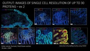
The interpretation of the tissue staining is challenging and requires knowledge in tissue morphology. Every analyzed tissue sample needs to be revised for quality control and exclusion of potential artefacts, necrotic regions etc. This important step is time consuming and requires expertise in tumor histology. Unfortunately, the researchers are often not aware of the importance of such quality control. Complexity is added by recent advances of classical staining techniques like immunohistochemistry (IHC), immunofluorescence (IF), FISH, SISH etc. Multiplexed methods are more and more commonly used in research projects and include antibody-based approaches such as Vectra Polaris or CODEX® or probe-based techniques such as in situ sequencing. Each technique comes with its own difficulties and limitations, which need to be identified and addressed in the corresponding tissue context.
At HistoOne, we have assembled an international team of pathologists with the specific aim to support the research community and companies with professional assistance. We cover a broad range of subspecialties such as dermatopathology, lung pathology, kidney pathology and gastrointestinal pathology as well as autopsy pathology. Importantly, our pathologists have experience in the interpretation of different staining techniques performed in research labs.
“Spatial biology is going to be able to help us dissect what is really important”
What pathology services do you provide and how do these support researchers involved in experimental/clinical studies?
We provide professional interpretation, evaluation and quantification of staining, considering tissue complexity and specific needs of an individual research project. We offer image ‘curation’ (i.e., removal of artefacts, necrosis, irrelevant areas etc.) and quality control as a preparation for any further image analysis.
However, our support goes beyond traditional pathology expertise, and also includes image analysis, with a specific focus on multiplex IHC/IF and spatial analysis.
Currently, we are working on the establishment of a complete staining pipeline as a service. We aim to offer our customers a full package, which turns their ideas into data, including each step from multiplex IHC/IF staining, to scanning, image analysis, data generation and interpretation.
Through multiplex IHC/IF, researchers now have a tool at hand to simultaneously analyze marker co-expression.
Why is multiplex IHC/IF an important tool for immunopathology?
I believe there are several important aspects. Through multiplex IHC/IF, researchers now have a tool at hand to simultaneously analyze marker co-expression. This is of major importance for proper identification of cell subsets with different phenotype, diverse functions and different activation status. For example, in our recent study, only by co-expression of two macrophage markers were we able to identity a specific macrophage subset of high predictive capacities in five tumor types.
Another important aspect is the possibility to apply spatial analysis. With the establishment of single-cell sequencing as a tool in molecular biology, the need to perform spatial mapping in situ has increased immensely. And thanks to Akoya, the field of spatial techniques is rapidly evolving. Still, very few groups possess and develop precise spatial analysis methods, as it requires a deep understanding in histology, image processing and data analysis.
Why did you choose the Vectra Polaris for your multiplex IHC/IF studies?
The Vectra Polaris system was installed at our department at Uppsala University several years ago. Although, in comparison to the CODEX system , the level of multiplexing may not seem that impressive (up to 9 markers), the Vectra Polaris is a very unique system when it comes to high throughput and reliability. In translational research, we usually have quite a large number of samples which need to be analyzed. On the other hand, after proper selection, there is typically only a very limited number of antibodies providing really good staining quality. Therefore, in practice, having 6-7 markers at one staining is more than enough for a properly designed translational research project. And don’t forget the complexity of data analysis, which comes after imaging! Vectra Polaris has very high throughput and requires only 15min to 2h for the scanning of a single slide. This is extremely important if you have many research groups using the machine.
Now, and with several years of the experience with Vectra Polaris, I can confirm that it was the proper decision to get this system installed.
What is the potential for multiplex IHC/IF for HistoOne and the services it can provide to researchers? How do you see it being used in the future?
From my experience, people often underestimate the efforts needed to establish perfect multiplex IHC/IF and generate reliable results. The Vectra Polaris system is not a limiting factor: as said, it is a very fine machine with a very robust and reliable workflow. However, there are two other factors, which require most of the efforts and time: wet-lab and image control/analysis. While the tissue scanning for one regular research project can be normally performed in one or a few days, the set-up of a proper multiplex marker ‘panel’ requires weeks of preparation. And the subsequent image/data analysis, if done properly, may take months, especially for unexperienced users.
To date, HistoOne can provide the service in image processing (quality control, analysis, post-imaging data processing) for the multiplex IHC/IF. We can also perform scanning on our system (in a frame of the collaboration with our university).
As referred to, currently, we are also establishing the wet-lab service. When this is done, we will be able to offer the complete pipeline from early multiplex panel establishment and tissue staining to scanning, image processing and data generation, saving researchers a lot of time, which they then can invest in other aspects of their project. HistoOne service will allow research groups to move forward and develop their ideas, instead of spending time optimizing staining and analysis of every marker of interest.
Our long-term goal is to develop AI-based pathology tools, aiming to support cancer diagnostic procedures and decision-making in clinical pathology.
What is the future vision for HistoOne?
Currently, we are working to establish the wet-lab service, to be able to offer the complete multiplex pipeline for researchers.
Apart from providing a service, HistoOne is deeply integrated into the research environment of Uppsala University, including biomarker discovery projects. Our recent finding, the signature of immune activation, is capable of predicting survival better than the state-of-art immune score in colon cancer and is prognostic also in other cancers with high mutational burden.
Our long-term goal is to develop AI-based pathology tools, aiming to support cancer diagnostic procedures and decision-making in clinical pathology. Recently, we have established a strategic agreement with Transport and Telecommunication Institute (TSI, Riga, Latvia), to combine our knowledge in pathology and their experience in the developing of AI-systems.
How can any interested researchers contact you about using your pathology services?
Any interested researchers can contact us by email at info@histo.one or simply drop me a message on LinkedIn.


