
Main Menu

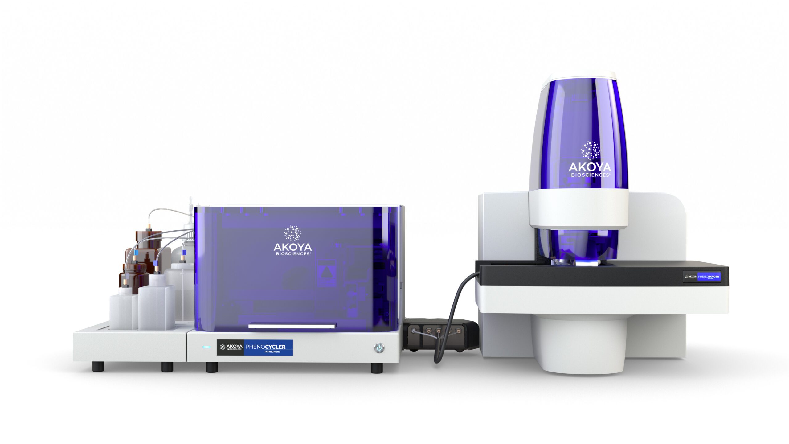
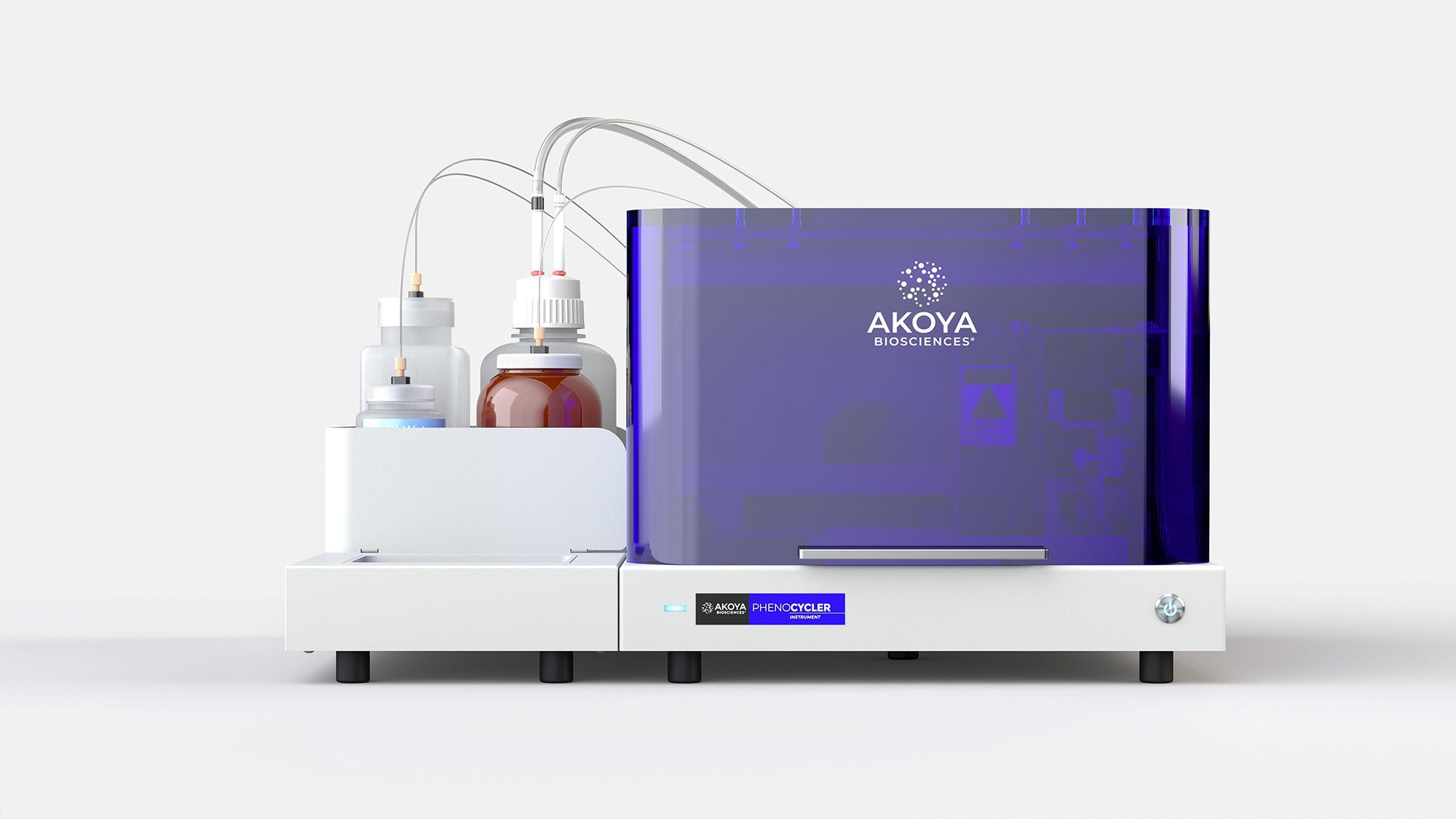
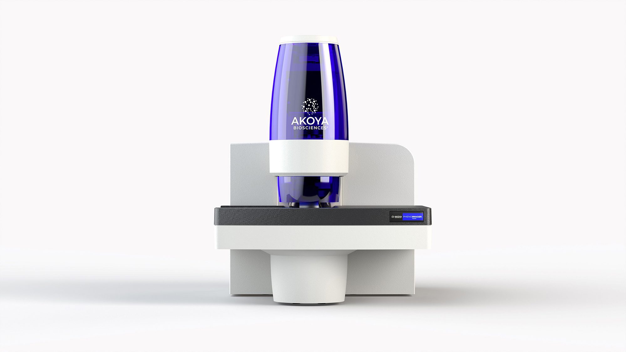
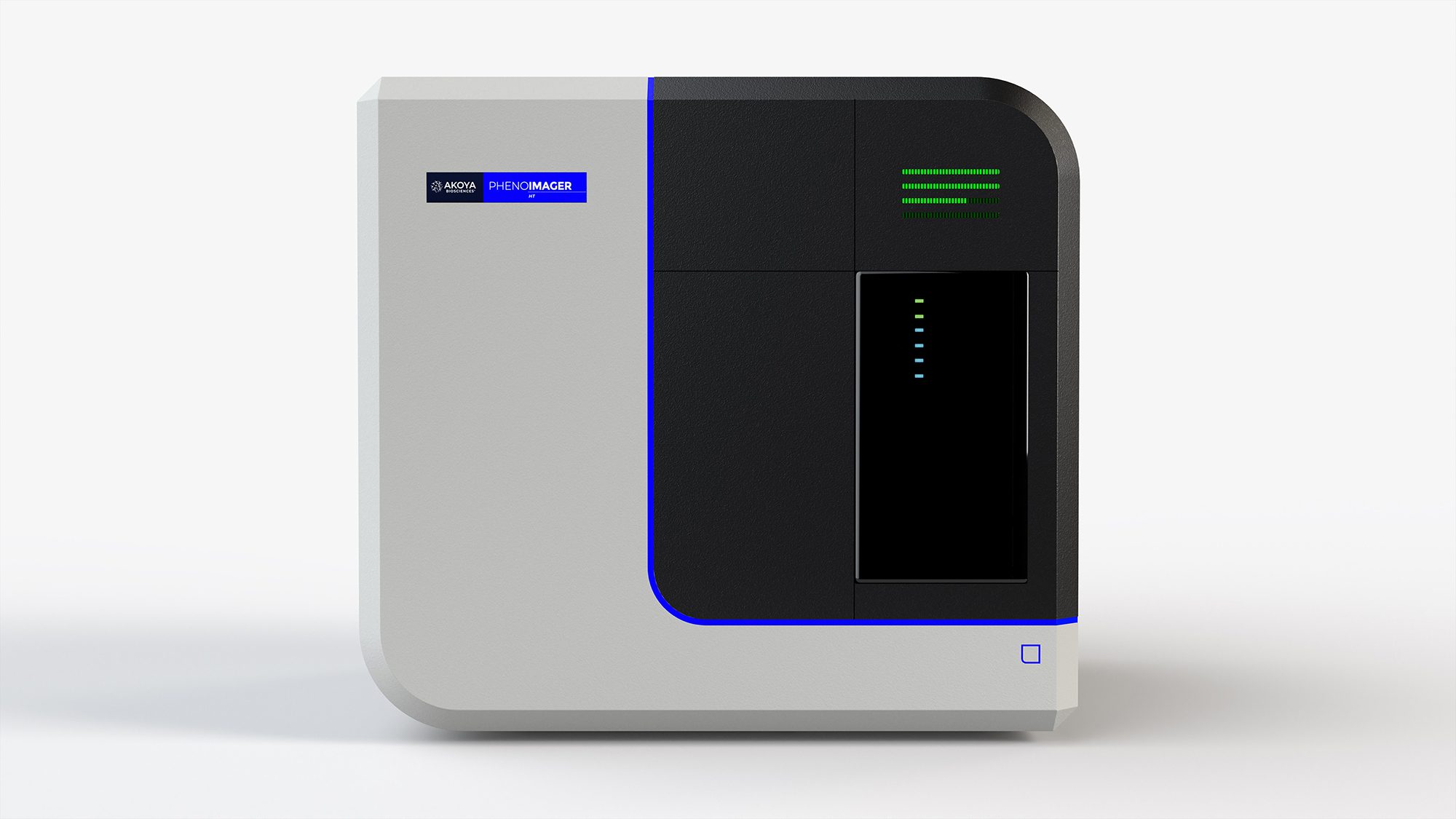
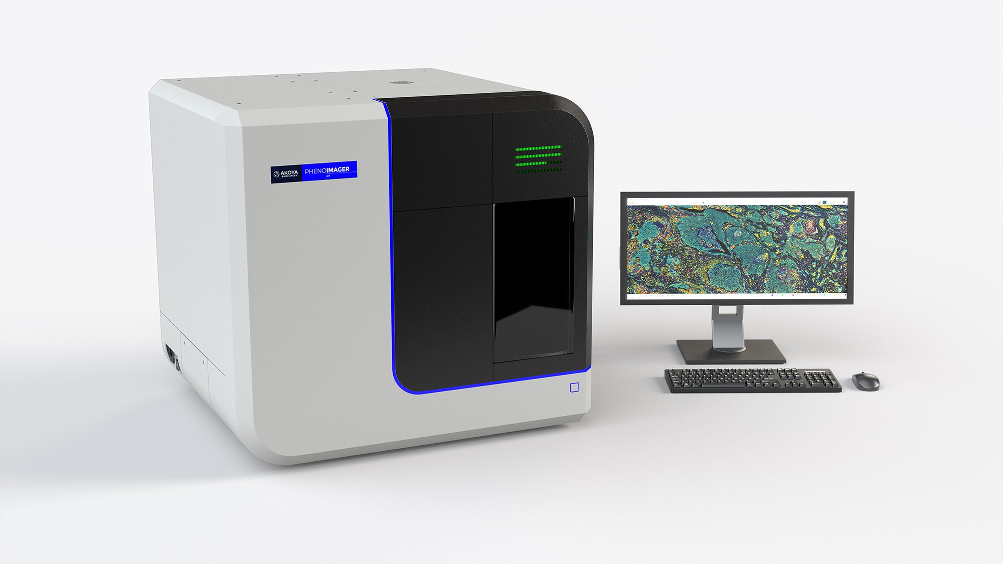
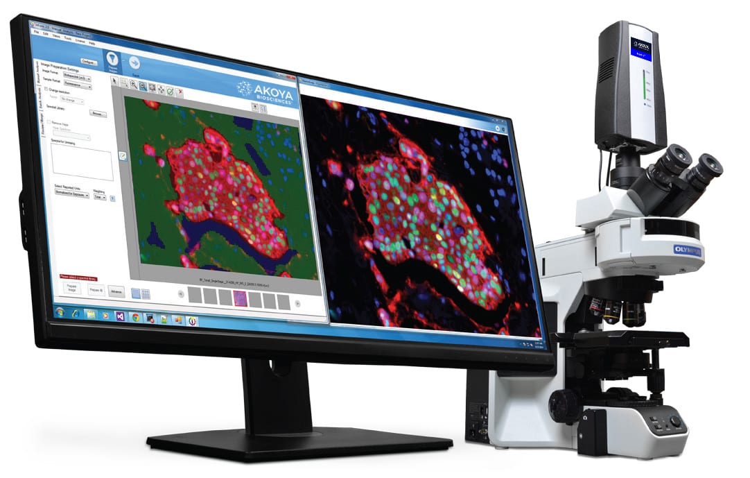
Images – Instrument


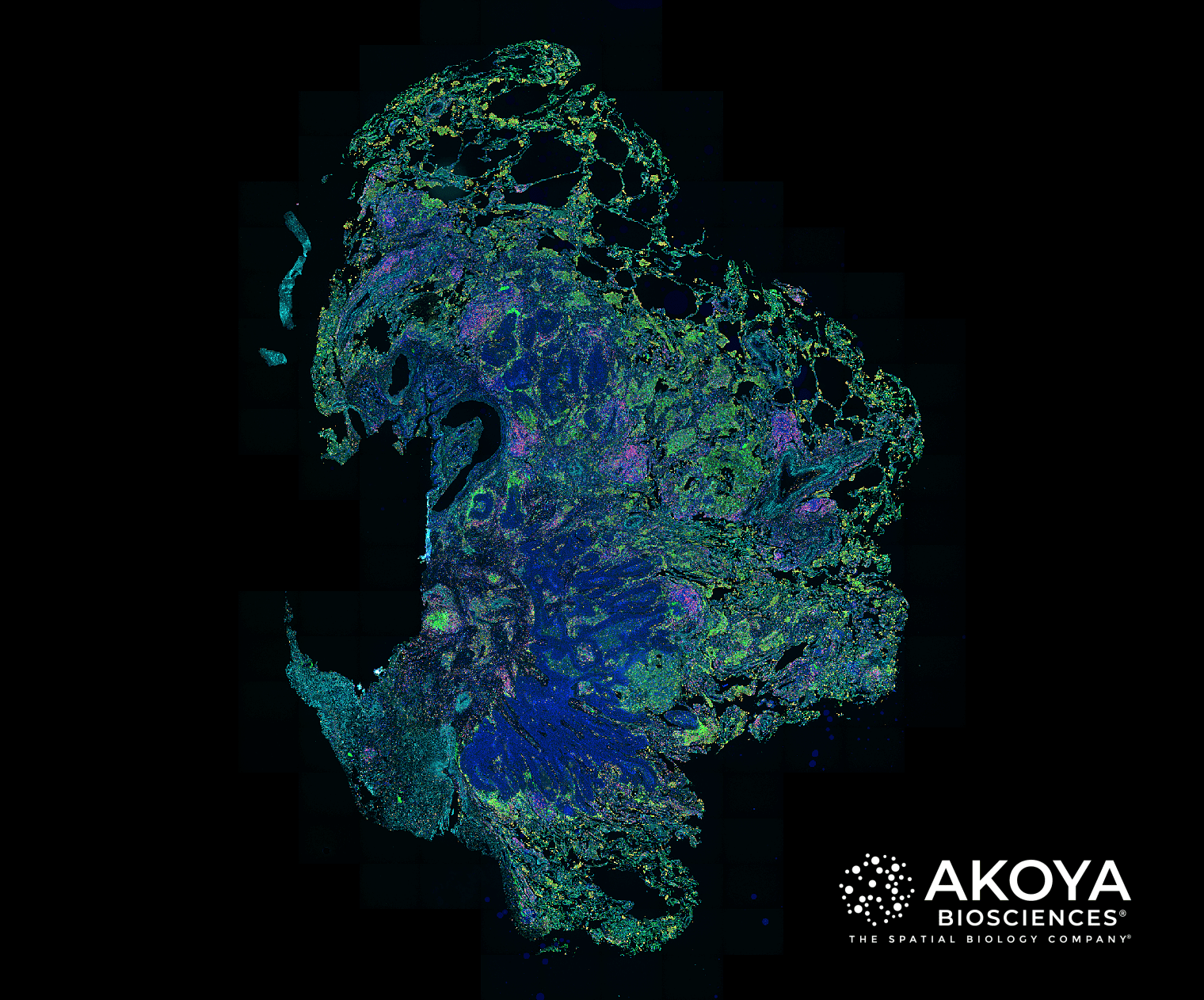
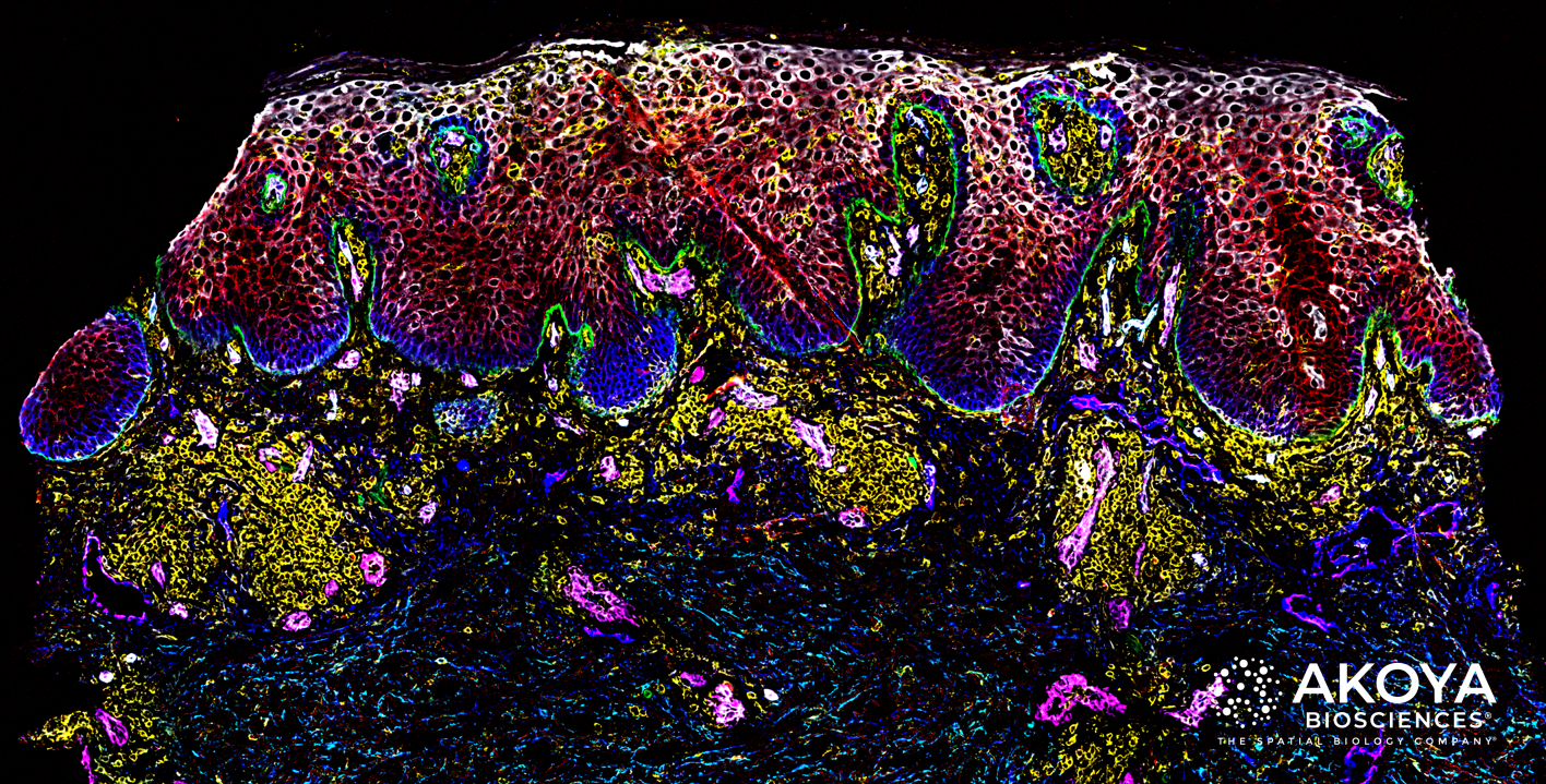
Images – Tissue


Logo with tagline


Logo without tagline


Press Releases
See our news releases page for a complete list of press releases.
Blog
See our blog page for a complete list of posts.
Case Studies
Infographics


Key Publications
 Synopsis: In a seminal multi-institutional study, published in JAMA Oncology in 2019 , a group of leading immuno-oncology experts determined that spatial phenotypic signatures, measured by multiplex immunofluorescence (mIF), outperformed other biomarker testing approaches in predicting response to anti-PD-1/PD-L1 treatments. The study was conducted by scientists at Johns Hopkins University, Yale University, Vanderbilt University, and Northwestern University.
The authors reviewed published data from more than 50 studies covering more than 10 types of cancer and over 8,000 patients. The mIF studies featured in this paper cite the use of Akoya’s Phenoptics™ platform.
 Synopsis: In a truly unique approach taken by scientists at Johns Hopkins University (JHU), Dr. Janis Taube, a leading pathology expert and Dr. Alex Szalay, a world-renowned astrophysicist, joined forces to solve the big data challenge in cancer biomarker discovery. They built a platform titled, AstroPath™ and applied celestial object mapping algorithms to the study of the tumor microenvironment, enabling rapid probabilistic studies of how tumor and immune cells organize and interact to influence treatment response.
The underlying image-capture technology for AstroPath is the PhenoImager HT (formerly Vectra® Polaris™), as part of an ongoing collaboration between Akoya and JHU. The result was the groundbreaking discovery of a spatial biomarker signature which is highly predictive of immunotherapy response in melanoma cases. The study was published in Science in 2021.
Videos


