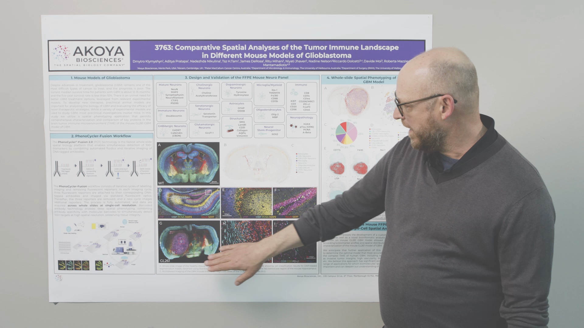
Welcome to part 2 of our AACR 2024 recap series. In this series, we highlight the great science and spatial biology technologies showcased at the AACR event in San Diego. Be sure to catch up on part 1 here if you missed it. In this second installment, we cover a poster demonstrating how neuroscientists can obtain deep insights into mouse brain cancer models using Akoya’s spatial biology solutions.
Spatial biology provides unparalleled insights into tumor immune microenvironment (TiME) complexity and unique opportunities to understand cancer-immune system interactions. Nowhere is this more critical than in tumors of the central nervous system, such as glioblastoma (GBM). A research collaboration between scientists at Akoya Biosciences, the Peter MacCallum Cancer Centre in Australia, and the University of Melbourne sought to utilize spatial phenotyping to characterize the immune landscape of GBM using an existing mouse model of the disease.
Findings from this work were shared in a poster presentation at the 2024 American Association for Cancer Research (AACR) annual meeting in San Diego.
Watch Video

Watch Akoya Technical Applications Scientist Fabrice Roegiers, PhD, present the poster, Comparative Spatial Analyses of the Tumor Immune Landscape in Different Mouse Models of Glioblastoma.
Research Focus: Addressing the Unmet Needs in Glioblastoma
GBM is a devastating disease and is the most commonly observed malignant tumor of the central nervous system. Despite advances in treatment, GBM remains one of the most challenging types of cancer to treat, and the prognosis for most patients is poor. The median survival time is about 12-15 months, and the five-year survival rate is less than 10%.
There is a clear and urgent unmet need for improved GBM treatment options, and comprehensive characterization of preclinical animal models using state-of-the-art technologies is needed to develop new therapies. Multiple forms of mouse models of GBM exist, ranging from xenografts to genetically engineered mice and syngenic murine models. In this study, the team used GL261, a chemically induced mouse model of glioblastoma, known to recapitulate histologic and biological characteristics of human GBM. Additionally, the GL261 model uses immunocompetent mice, so it is suitable for analyzing GBM tumor immunology and immunotherapies.
For this study, the research team performed spatial phenotyping – using Akoya’s spatial biology solutions – to characterize and compare critical proteins in the brain TiME comprehensively with the help of this model.
Technology Spotlight: Whole Slide Imaging Offers New Perspective on GBM
The team designed and validated an 43-plex antibody panel comprised of protein markers spanning numerous cell types including neuronal and glial cells, immune populations, structural features, and neuropathological markers. To fully map the tumor immune landscape of mouse GBM, they used the PhenoCycler-Fusion 2.0, which allows scientists to capture whole-slide, highly multiplexed tissue images. This approach allows for an entire tissue section of the brain to be captured as a single image while also achieving single-cell resolution. This facilitates the detection of global changes in cell distribution and histology while concurrently quantifying the presence of biomarkers in individual cells.

Key Findings: Spatial Phenotyping of Mouse GBM Model Provides New Depths of Understanding
- Using the newly established Mouse Neuro Panel, spatial mapping of wild-type and GBM tissue sections revealed unique distributions of cell types and biomarker accumulation.
- The GBM tissue spatial map and accompanying t-SNE plot showed distinct phenotype clusters quantified into 14 cell types.
- Looking further at the expression of individual markers, functional specialization was shown across the entire GL261 brain tissue section. Specifically, biomarker staining revealed immune cell populations infiltrating the tumor mass and displacement of neuronal and glial cell types.
Download the full poster in PDF format for a closer look.
Conclusions: Mouse Panel Yields New Paths to Understanding GBM
The data presented in this poster represent the successful development of a custom antibody panel, imaging workflow, and a novel bioinformatic analysis method. Using this workflow and the PhenoCycler-Fusion 2.0 enabled exploration of the TiME of the mouse GL261 GBM model with never-before-achieved resolution. Through 43 biomarker profiles and their respective spatial distributions, the research team was able to characterize the distribution of 14 cell populations in healthy and GBM-affected brain tissue.
Future application of this panel will aid in determining optimal mouse models that most accurately recapitulate the complex TiME of human GBM, including key features such as invasive tumor margins, high vascularity, blood-brain barrier, etc. Ultimately, spatial phenotyping data will support the development of next-generation therapeutics for treating GBM.
For more information:
- See how the PhenoCycler-Fusion 2.0 solution is helping provide insights into complex biological systems and gain a better understanding of cellular microenvironments.
- Watch the on-demand recording of our AACR Spotlight Theater presentation, “Spatial Biology 2.0: Spatial Insights & Precision Medicine at Unprecedented Scale”.
- Catch up on part 1 of the AACR 2024 Recap Series.


