
A ZEISS Microscopy User Story
When our immune systems identify invaders – such as parasites, bacteria or viruses – it can spark inflammation, which brings fluid and immune cells to the area of infection. While this can be very effective in battling infection, it also has collateral damage on nearby healthy tissues. In our daily lives, just by eating or breathing, we are constantly exposing our immune systems to microbes and particulate matter that can elicit low level inflammatory responses. The impact of these events over time is not well understood.
Mononuclear phagocyte cells (MPs) are myeloid cells and key regulators in the inflammatory process. They are present in every tissue and are very diverse with multiple origins.
One of the goals in Prof. Andreas Schlitzer’s lab at the Life and Medical Sciences Institute (LIMES) of the University of Bonn (Germany) is to understand the distribution, function and origin of MPs in healthy tissues and during acute or chronic inflammation. We spoke with Dr. David Bejarano, a postdoctoral researcher in this lab, about how he is using a multiplex tissue microscopy system to characterize the physical locations and relationships of immune cells in these tissues.
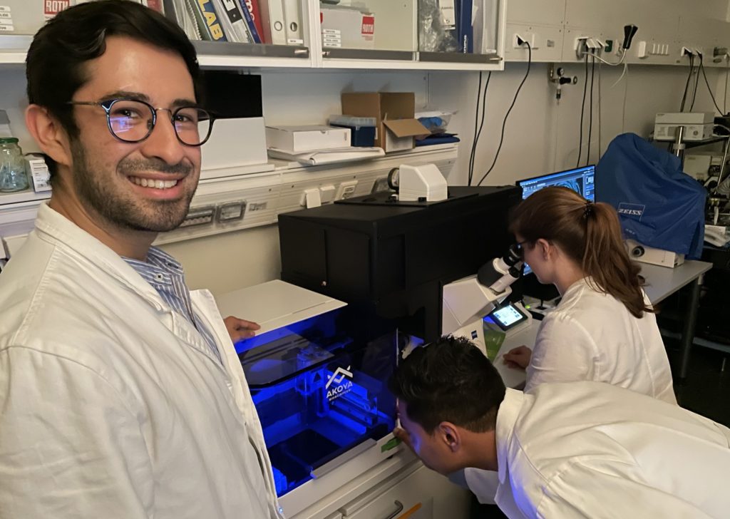
Dr. David Bejarano (left) with graduate students Sumit Sheoran and Hannah Theobald
When did you first become interested in multiplex tissue microscopy?
When we read this paper from Dr. Garry Nolan’s group at Stanford University (USA), we got very excited because they presented a very solid and elegant strategy to detect more than 40 markers/antibodies, simultaneously, which they called CODEX Multiplexed Imaging. In 2019, we learned that Akoya Biosciences was creating a commercialized system and we decided to establish it in our group. An important element was selecting the proper microscope that could integrate with the Akoya Biosciences solution. Although there were several options in the market, the ZEISS Axio Observer microscope offered the best sensitivity and an optimal speed of acquisition. We can acquire the full left murine lung stained with 40 antibodies in less than 32 hours.
How has the Axio Observer microscope with CODEX workflow changed your research?
Before acquiring the Axio Observer microscope and the CODEX multiplex system, our findings were mainly based on high dimensional flow cytometry and single-cell transcriptomics. Although both approaches allow a deep understanding of immune responses at a single cell level, they lack spatial resolution and context.
However, cells do not live in isolation but are part of an intricate network of interactions and interdependence. We realized we needed to find a way to study the complexity of the innate immune system while keeping the context of the tissue.


3 µm section of a formalin fixed paraffin-embedded human lung. Section was stained with a 14 CODEX antibody panel. Image shows some essential markers to distinguish mononuclear phagocytes (CD45 in gray, CD68 in red, HLA-DR in green, CD11c in magenta). To delineate major anatomical structures, such as bronchioles and blood vessels, Pancytokeratin (yellow) and CD31 (cyan) are shown. Blue signals correspond to DAPI nuclear staining. Acquisition was done with a ZEISS Axio Observer microscope and processing with the Akoya CODEX Processor.
The incorporation of a highly multiplexed imaging technique that allows visualizing at a single-cell level more than 40 cell markers in record time without compromising tissue morphology or image quality has brought our research to another level of understanding. With CODEX we want to (and have been able to) visualize major tissue morphological changes induced by different conditions, localize cells of interest, characterize the surrounding of these cells, look at their interaction partners and determine how the local environment shapes the function of a cell. It has been fascinating to see how using the CODEX technology with our Axio Observer microscope adds a whole new dimension/parameter to the insights we are able to obtain.
These datasets are just really amazing windows into cell biology!


1 µm section of a formalin fixed paraffin-embedded human lymph node. Section was stained with a 14 CODEX antibody panel. Image shows some essential markers to distinguish mononuclear phagocytes (CD68 in red, HLA-DR in green, CD11c in magenta). To delineate major anatomical structures, such as blood vessels, CD31 is shown in cyan. Proliferation marker Ki67 (yellow) delineates germinal centers. Blue signals correspond to DAPI nuclear staining. Acquisition was done with a ZEISS Axio Observer microscope and processing with the Akoya CODEX Processor.
Can you provide a teaser about your initial findings?
We have been using the system for a year and we keep finding ways to pull out more and better information through our analysis pipeline. Basically almost all of our current projects are profiting from the CODEX multiplex tissue microscopy system workflow!
We have a couple of projects that have started with an exploration of the spatial distribution and diversity of myeloid cells using CODEX. For example, we have looked at macrophages and dendritic cells in the longitudinal axis of the murine large intestine and found that there is a gradient in their distribution. We are currently focusing on the characterization of macrophage populations that we observe in the different layers of the intestinal wall and would like to use spatial-omics in fate mapping mouse models to track their origin.

5 µm section of a formalin fixed paraffin-embedded mouse small intestine swiss roll. Section was stained with a 14 CODEX antibody panel designed to visualize intestinal stem and enteroendocrine cells (Lysozyme in red, villin in green, MHCII in gray, vimentin in cyan, Ki67 in magenta, and Olfm4 in yellow). Blue signals correspond to DAPI nuclear staining. Acquisition was done with a ZEISS Axio Observer microscope and processing with the Akoya CODEX Processor.
In another project, we are studying the effect of low-grade inflammation in the lung which as been induced by exposure to environmental fungal polysaccharides on subsequent acute or chronic inflammatory episodes. After stimulation, there is a rapid and pronounced influx of a subpopulation of alveolar macrophages that have a pro-inflammatory profile that is kept for several weeks and enhances the response to subsequent challenges. Interestingly, when we induce pulmonary fibrosis in mice that had been stimulated, the disease progression is slower and survival rates improve. Besides doing a detailed characterization of these cells and the genes controlling this phenotype, we used a 40-plex CODEX antibody panel to visualize the lung architecture and determine if major structural changes occur.


3 µm frozen section of the left mouse lung 3 days after intranasal stimulation with LPS. Section was stained with a 37 CODEX antibody panel. Image shows the canonical markers of mononuclear phagocytes (CD11c in red, MHCII in green, F4/80 in cyan, CD11b in magenta). To delineate major anatomical structures, such as lung airways and blood vessels, EpCAM (yellow) and SMA (gray) are displayed. Blue signals correspond to DAPI nuclear staining. Acquisition was done with a ZEISS Axio Observer microscope and processing with the Akoya CODEX Processor.
In agreement with our flow cytometry analyses, the images showed an increase of monocytes and monocyte-derived alveolar macrophages. They also revealed the absence of major structural changes. Nonetheless, in the aerial spaces, such as bronchioles, infiltrates of cells were observed. These infiltrates were composed predominantly of the macrophage/monocyte populations we had identified, but also other myeloid cells and even lymphoid cells were present. We are currently determining how long these cell infiltrates last and if they are abolished when the master regulators of our phenotype are genetically ablated in the mice. This is a nice example of how adding spatial analyses broadens our understanding of the immune response.
Where do you see this project going within the next year?
There are many goals that we have in mind. We would like to determine the influence of the cellular niche on the function of a cell. There are multiple possible approaches, one of them is to include markers in our CODEX antibody panels to visualize immune activation and cellular metabolism. Furthermore, we would like to exploit the high dimensionality of the technique to study the diversity of myeloid cells in multiple tissues and identify non-described tissue-specific populations. There is so much we still don’t understand and we see great potential in leveraging such findings to eventually lead to new and better targeted treatments for multiple health conditions.
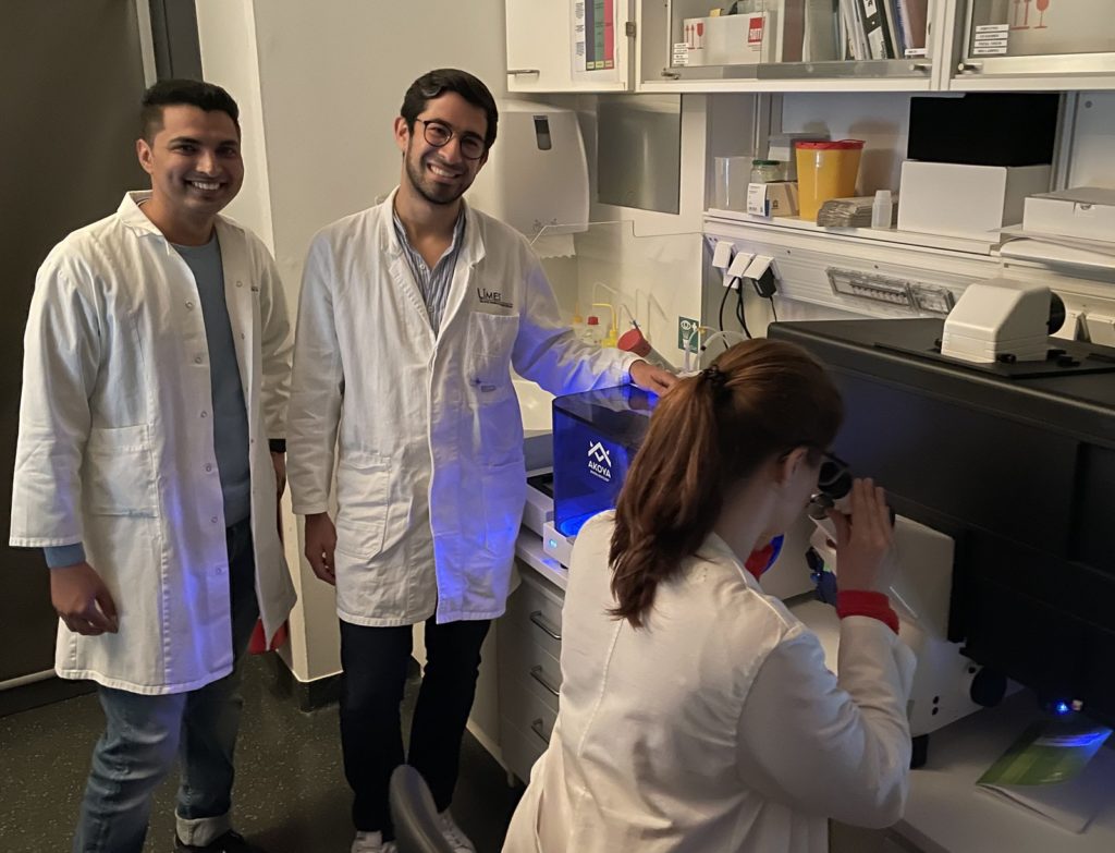
Dr. David Bejarano (center) with Sumit Sheoran and Hannah Theobald
Our most ambitious goal is to combine and correlate our CODEX data with single-cell RNA sequencing and high dimensional flow cytometry data in multiple murine and human tissues to create a comprehensive cellular atlas of myeloid cells that improves their identification and allows understanding of their distribution, localization and function. All four of our users of the CODEX multiplex tissue microscopy system from our group and our bioinformatician have teamed up with another group of bioinformaticians from the LIMES Institute. Besides the experimental work, we are dealing with the construction of a pipeline and also with a massive amount of data! But we hope that in a year we will be able to share our results.
This blog was originally published as a ZEISS Microscopy User Story. To read more articles about applications for ZEISS microscopes, visit their blog page.


