Webinar

webinar thumbnail jcool czi

Single-Cell, Spatial Phenotyping: Fueling the Next Wave of Discovery Biology

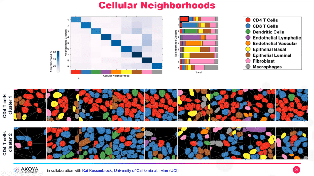
In this webinar Dr. Jonah Cool, Program Officer for Single-Cell Biology at the Chan Zuckerberg Initiative, provides an overview of single-cell efforts at CZI; and Dr. Oliver Braubach, Head of Research Applications at Akoya Biosciences, introduces the CODEX solution and shares case studies on how single-cell, spatial phenotyping applications can push the boundaries of discovery biology in human health and disease.

Single-Cell, Spatial Phenotyping: Fueling the Next Wave of Discovery Biology Read More »

webinar thumbnail jcvillasboas immunome

The Cancer Immunome Project: Adding the Spatial Dimension

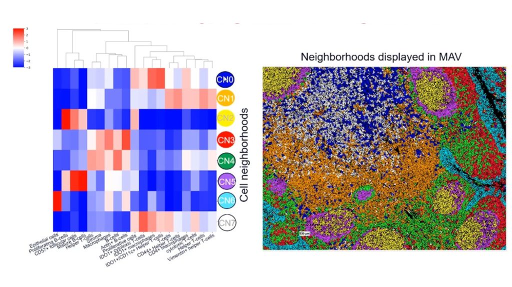
Immune checkpoint inhibitors are revolutionizing cancer therapy for many. But robust biomarkers are needed to predict which patients will likely benefit from these therapies. A team of investigators at the Mayo Clinic is trying to close this gap by developing a catalog of the human immune system in cancer patients, appropriately called “The Cancer Immunome Project. By analyzing the spatial architecture of FFPE tumor sections across the entire tumor microenvironment, the team is generating a comprehensive map of the cancer immunome. In this webcast, Dr. JC Villasboas shares how his team is using the CODEX® System to deepen our knowledge about the composition, architecture and interactions of the tumor microenvironment.

The Cancer Immunome Project: Adding the Spatial Dimension Read More »

webinar thumbnail spatial bio europe 2

Spatial Biology Europe 2021: Broaching the Next Frontier of Cellular Identification with Single-Cell, Spatial Phenotyping

Hear from Oliver Braubach, Ph.D., Senior Manager, Applications at Akoya Biosciences on the importance of single cell resolution in spatial biology , how to discover single cell contextual phenotypes and the many single-cell, spatial phenotyping applications.

Spatial Biology Europe 2021: Broaching the Next Frontier of Cellular Identification with Single-Cell, Spatial Phenotyping Read More »

webinar thumbnail spatial bio europe 1

Spatial Biology Europe 2021: A Spatial Biology Approach For Biomarker Discovery and Validation in Immuno-Oncology

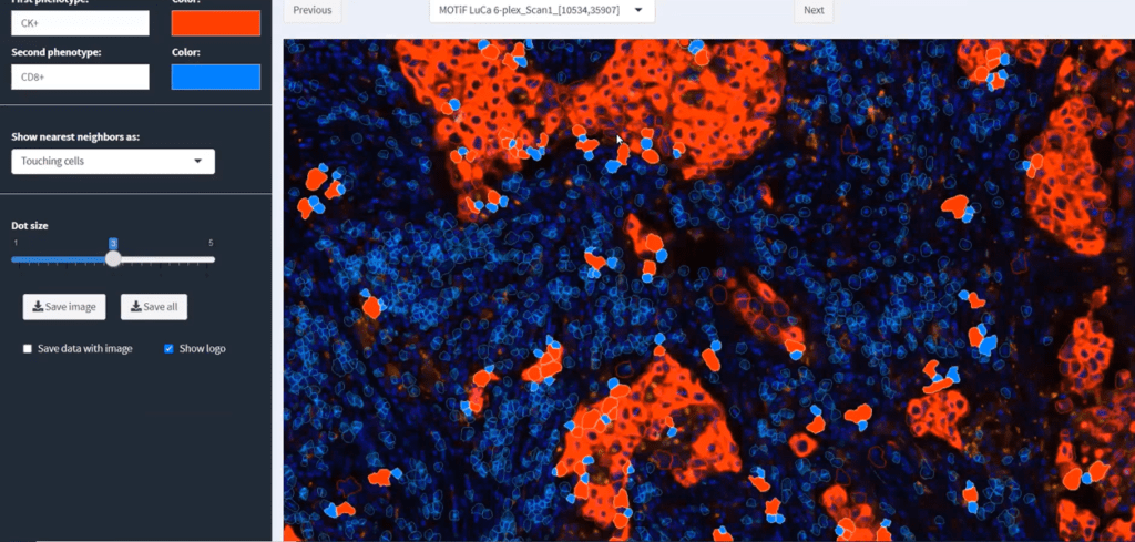
In this presentation, from the 2021 Spatial Biology Europe Congress, Gavin Gordon, Ph.D., Vice President, Clinical Market Development from Akoya Biosciences describes evolving biomarker strategies in immuno-oncology and how multiplexed immunofluorescence has been associated with improved predictive performance. Dr Sebastian Marwitz, ARCN Principal Investigator, Pathology from Research Center Borstel – Leibniz Lung Center, describes how multiplexed IHC has been used for the spatial profiling in lung disease in hypotheses-driven analyses at a cohort level using FFPE patient samples.

Spatial Biology Europe 2021: A Spatial Biology Approach For Biomarker Discovery and Validation in Immuno-Oncology Read More »

webinar thumbnail jgalon

Spatial Phenotyping of the Tumor Microenvironment: Implications for Immunotherapy

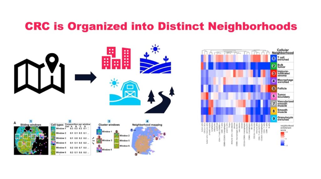
Recent studies strongly suggest the importance of determining a patient’s Immunoscore as well as the need for a more comprehensive understanding, both spatially and functionally, of the simultaneous presence of multiple immune cell types within the TME. Immune contexture parameters, including the Immunoscore, have a prognostic, predictive, and mechanistic value. Once identified, the key immune elements should be translated into clinically feasible treatment protocols, integrating with the field of immunopathology. In this webinar, Dr. Jerome Galon describes how a spatial phenotyping approach has supported the development of Immunoscore to estimate the prognosis of colorectal cancer patients

Spatial Phenotyping of the Tumor Microenvironment: Implications for Immunotherapy Read More »

webinar thumbnail spatial multiomics

Spatial Multiomics Webinar Series

In this multi-part webinar series, our expert speakers review analytical frameworks and algorithms to integrate imaging-based single-cell spatial phenotyping data with complementary transcriptomic and genomic datasets. High-plex cell phenotyping methods like single-cell RNA-seq capture the deep cellular heterogeneity of samples, but cell behavior is a function of all that surrounds it. Imaging-based spatial phenotyping platforms enable researchers to visualize and analyze cell diversity, interactive networks, and cellular behavior across whole tissue sections. Both types of data have complementary features, which give researchers the ability to merge information about a cell’s proteome and transcriptome with its single-cell, spatial context. This webinar series highlights the latest advances driving integrative multiomic analysis.

Spatial Multiomics Webinar Series Read More »

webinar thumbnail biocompare sTILs PD L1

Using Multiplex Immunofluorescence to Better Quantify Treatment-Related Changes in sTILs/PD-L1

H&E stromal tumor-infiltrating lymphocyte (sTIL) score and programmed death ligand 1 (PD-L1) SP142 immunohistochemistry assay are prognostic and predictive in early-stage breast cancer but are operator-dependent and may have insufficient precision to characterize dynamic changes in sTILs/PD-L1 in the context of clinical research. In this webinar, the presenters discuss how multiplex immunofluorescence (mIF) combined with statistical modeling can be used to estimate dynamic changes in sTIL score, PD-L1 expression, and other immune variables from a single paraffin-embedded slide, thus enabling comprehensive characterization of activity of novel immunotherapy agents.

Using Multiplex Immunofluorescence to Better Quantify Treatment-Related Changes in sTILs/PD-L1 Read More »

webinar thumbnail simulated he

R&D Sneak Peek | Opal Multiplex and Simulated H&E on The Same Tissue Section

In today’s clinical research laboratories, available tissue samples are both smaller in size and more in demand for a variety of assays to help researchers make informed decisions and spatial discoveries. The very cornerstone of tissue histopathology – the H&E stain – is the first crucial step to help map out the morphological landscape. Here, Akoya Biosciences describes a technique in which a tissue sample is stained with a modified H&E stain prior to the application of an Opal Multiplex assay on the same slide, as a screening tool and efficient and effective way to maximize your precious samples.

R&D Sneak Peek | Opal Multiplex and Simulated H&E on The Same Tissue Section Read More »

webinar thumbnail columbia science

Validating a Multiplex Immunofluorescence Workflow to Improve Stratification of High-Risk, Early-Stage Melanoma Samples

Dr. Yvonne Saenger, from the Columbia University Herbert Irving Comprehensive Cancer Center, discusses how she used a validated quantitative multiplex immunofluroescence imaging workflow to identify individuals who would respond to adjuvant immunotherapy, better stratifying them into high-risk versus low-risk stage II melanoma.

Validating a Multiplex Immunofluorescence Workflow to Improve Stratification of High-Risk, Early-Stage Melanoma Samples Read More »