Webinar

webinar thumbnail spatial bio europe 1

Spatial Biology Europe 2021: A Spatial Biology Approach For Biomarker Discovery and Validation in Immuno-Oncology

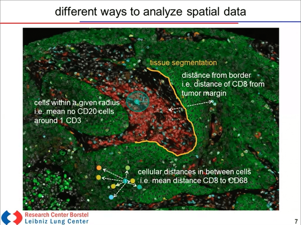
In this presentation, from the 2021 Spatial Biology Europe Congress, Gavin Gordon, Ph.D., Vice President, Clinical Market Development from Akoya Biosciences describes evolving biomarker strategies in immuno-oncology and how multiplexed immunofluorescence has been associated with improved predictive performance. Dr Sebastian Marwitz, ARCN Principal Investigator, Pathology from Research Center Borstel – Leibniz Lung Center, describes how multiplexed IHC has been used for the spatial profiling in lung disease in hypotheses-driven analyses at a cohort level using FFPE patient samples.

Spatial Biology Europe 2021: A Spatial Biology Approach For Biomarker Discovery and Validation in Immuno-Oncology Read More »

webinar thumbnail jgalon

Spatial Phenotyping of the Tumor Microenvironment: Implications for Immunotherapy

Recent studies strongly suggest the importance of determining a patient’s Immunoscore as well as the need for a more comprehensive understanding, both spatially and functionally, of the simultaneous presence of multiple immune cell types within the TME. Immune contexture parameters, including the Immunoscore, have a prognostic, predictive, and mechanistic value. Once identified, the key immune elements should be translated into clinically feasible treatment protocols, integrating with the field of immunopathology. In this webinar, Dr. Jerome Galon describes how a spatial phenotyping approach has supported the development of Immunoscore to estimate the prognosis of colorectal cancer patients

Spatial Phenotyping of the Tumor Microenvironment: Implications for Immunotherapy Read More »

webinar thumbnail spatial multiomics

Spatial Multiomics Webinar Series

In this multi-part webinar series, our expert speakers review analytical frameworks and algorithms to integrate imaging-based single-cell spatial phenotyping data with complementary transcriptomic and genomic datasets. High-plex cell phenotyping methods like single-cell RNA-seq capture the deep cellular heterogeneity of samples, but cell behavior is a function of all that surrounds it. Imaging-based spatial phenotyping platforms enable researchers to visualize and analyze cell diversity, interactive networks, and cellular behavior across whole tissue sections. Both types of data have complementary features, which give researchers the ability to merge information about a cell’s proteome and transcriptome with its single-cell, spatial context. This webinar series highlights the latest advances driving integrative multiomic analysis.

Spatial Multiomics Webinar Series Read More »

webinar thumbnail biocompare sTILs PD L1

Using Multiplex Immunofluorescence to Better Quantify Treatment-Related Changes in sTILs/PD-L1

H&E stromal tumor-infiltrating lymphocyte (sTIL) score and programmed death ligand 1 (PD-L1) SP142 immunohistochemistry assay are prognostic and predictive in early-stage breast cancer but are operator-dependent and may have insufficient precision to characterize dynamic changes in sTILs/PD-L1 in the context of clinical research. In this webinar, the presenters discuss how multiplex immunofluorescence (mIF) combined with statistical modeling can be used to estimate dynamic changes in sTIL score, PD-L1 expression, and other immune variables from a single paraffin-embedded slide, thus enabling comprehensive characterization of activity of novel immunotherapy agents.

Using Multiplex Immunofluorescence to Better Quantify Treatment-Related Changes in sTILs/PD-L1 Read More »

webinar thumbnail simulated he

R&D Sneak Peek | Opal Multiplex and Simulated H&E on The Same Tissue Section

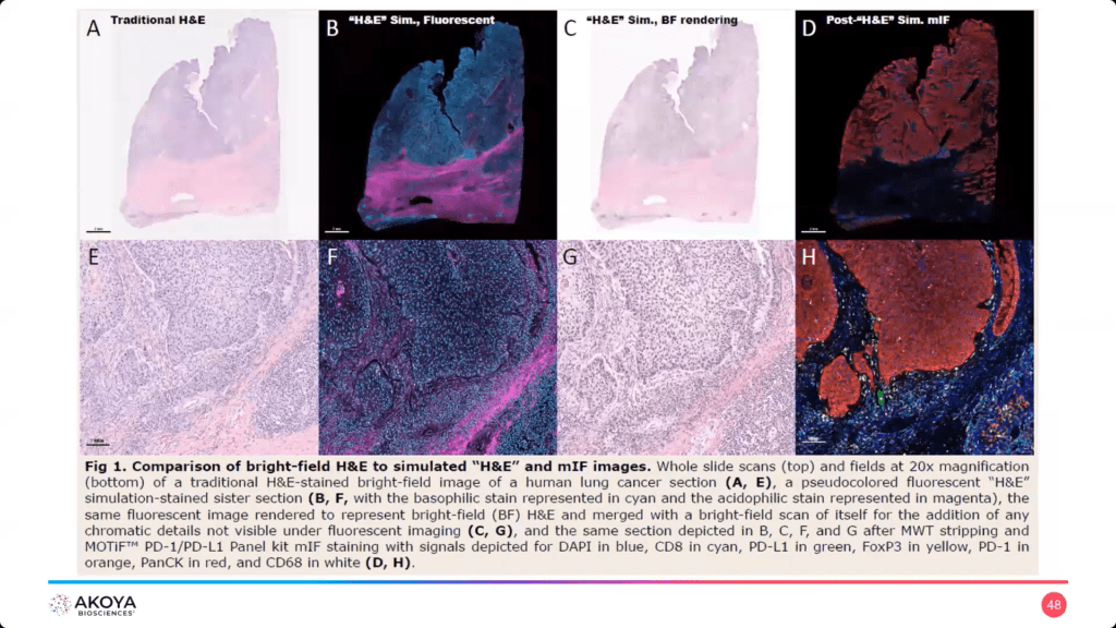
In today’s clinical research laboratories, available tissue samples are both smaller in size and more in demand for a variety of assays to help researchers make informed decisions and spatial discoveries. The very cornerstone of tissue histopathology – the H&E stain – is the first crucial step to help map out the morphological landscape. Here, Akoya Biosciences describes a technique in which a tissue sample is stained with a modified H&E stain prior to the application of an Opal Multiplex assay on the same slide, as a screening tool and efficient and effective way to maximize your precious samples.

R&D Sneak Peek | Opal Multiplex and Simulated H&E on The Same Tissue Section Read More »

webinar thumbnail columbia science

Validating a Multiplex Immunofluorescence Workflow to Improve Stratification of High-Risk, Early-Stage Melanoma Samples

Dr. Yvonne Saenger, from the Columbia University Herbert Irving Comprehensive Cancer Center, discusses how she used a validated quantitative multiplex immunofluroescence imaging workflow to identify individuals who would respond to adjuvant immunotherapy, better stratifying them into high-risk versus low-risk stage II melanoma.

Validating a Multiplex Immunofluorescence Workflow to Improve Stratification of High-Risk, Early-Stage Melanoma Samples Read More »

webinar thumbnail human cell atlas

Human Cell Atlas: A Spatially Resolved Map of Human Breast Tissue

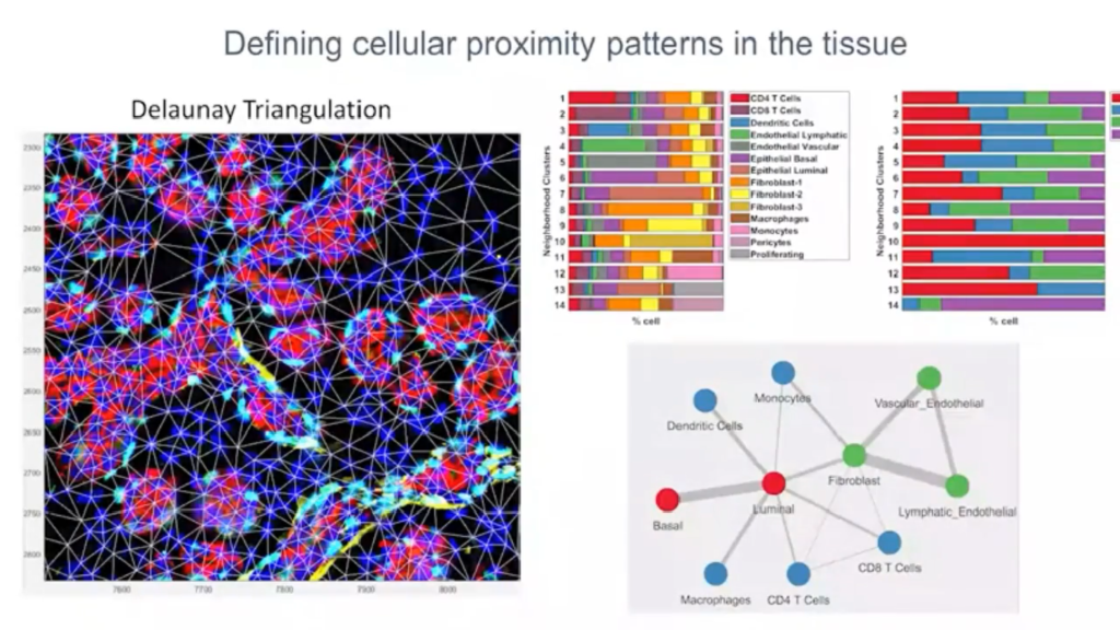
In this webcast, Dr. Kai Kessenbrock explains how spatial phenotyping can enhance the biological insights from Human Cell Atlas initiatives. Using the Human Breast Cell Atlas project as an example, Dr. Kessenbrock discusses how his team discovered unique cellular niches within the breast tissue microenvironment and how their spatial proximity gives us a more comprehensive view of the biology underlying each sample.

Human Cell Atlas: A Spatially Resolved Map of Human Breast Tissue Read More »

SITC Thumbnail v2

T.I.M.E and Space to Predict Response in Immuno-Oncology

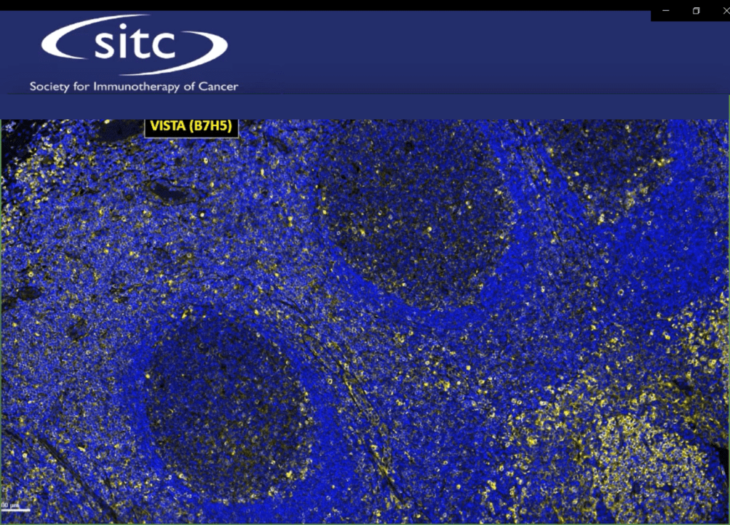
In this talk, Dr. Alexander “Sandy” Borowsky will share preliminary results of an ongoing biomarker discovery study on breast cancer cohorts, including those participating in the I-SPY 2 trial, and how the in situ analysis of the tumor microenvironment is critical to understanding immunotherapy response. The I-SPY 2 trial: Neoadjuvant and Personalized Adaptive Novel Agents to Treat Breast Cancer was launched by UCSF in collaboration with a private-public partnership to improve efficiency of breast cancer clinical trials and streamline the development of new drugs. Breast cancers, overall, infrequently respond to immunotherapy. And immunotherapies, which harness the immune system to fight cancer, are being used in the early high-risk treatment setting. However, individual tumors show dramatic variation in patterns of the tumor immune microenvironment (TIME) with both immune “cold” and inflamed phenotypes. We hypothesized that the TIME pattern might predict responses to immunotherapy and/or prognosis/outcomes. We examined the TIME of high-risk invasive breast cancer and high-risk non-invasive ductal carcinoma in situ (DCIS), hypothesizing that immune evasion might be one of the critical steps in progression of DCIS to invasive carcinoma. Leveraging current clinical trials, we have performed gene expression analyses (bulk) and multiplexed-immunohistochemistry (mIHC). We have optimized two 7-color assay panels using the Opal detection and artificial intelligence-assisted image analysis to provide specific cell segmented assignment of location and applied proximity analyses for cell-cell interaction assessment. These data show distinct differences in the TIME and reveal predictions of early response.

T.I.M.E and Space to Predict Response in Immuno-Oncology Read More »

Webinar Thumbnail

How Autofluorescence Reduction can Improve Your CODEX Workflow

Autofluorescence is a well-known problem associated with multiplex imaging applications, including PhenoCycler (formerly CODEX). We demonstrate a simple autofluorescence reduction method that integrates readily into the CODEX workflow. The protocol improves CODEX imaging data and streamlines antibody panel design by enabling more reliable use of low wavelength fluorophores, such as Alexa 488.

How Autofluorescence Reduction can Improve Your CODEX Workflow Read More »

webinar thumbnail covid multiorgan

COVID-19 Multiorgan Study: Spatial Phenotyping of Immune Cell Subsets in Patients with Lethal COVID-19

In this webinar, researchers from the University of Birmingham and the University of Limerick describe how they used multiplexed imaging and single-cell analysis to study the immune response to the SARS-CoV-2 virus across multiple organ systems. Deep immune phenotyping of COVID-19 tissue sections can reveal how the spatial and structural relationships between the virus and different cell types can influence disease pathology and progression.

COVID-19 Multiorgan Study: Spatial Phenotyping of Immune Cell Subsets in Patients with Lethal COVID-19 Read More »